EIZO product catalog
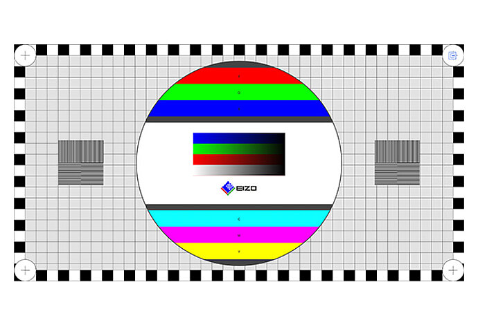
Test the properties and parameters of your monitor quickly and easily with software developed in-house at EIZO.
The ColorNavigator software is used to simply and accurately calibrate ColorEdge screens.
EIZO software allows you to easily manage usage and configuration for a single screen or multi-monitor setup. Windows and macOS operating systems are supported.
The EIZO software is capable of complete quality management – from calibration through asset management to acceptance and constancy testing.
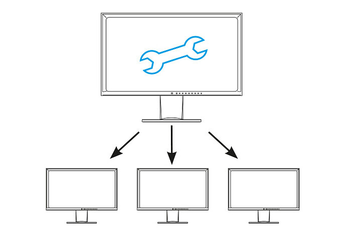
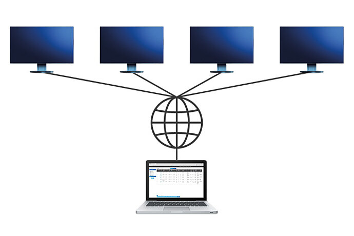
EIZO Monitor Configurator facilitates cross-network installation when monitors in a company need to have the same settings.
Quick Color Match is a software that makes complex color management workflow much easier for home inkjet printers.
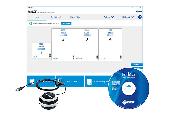
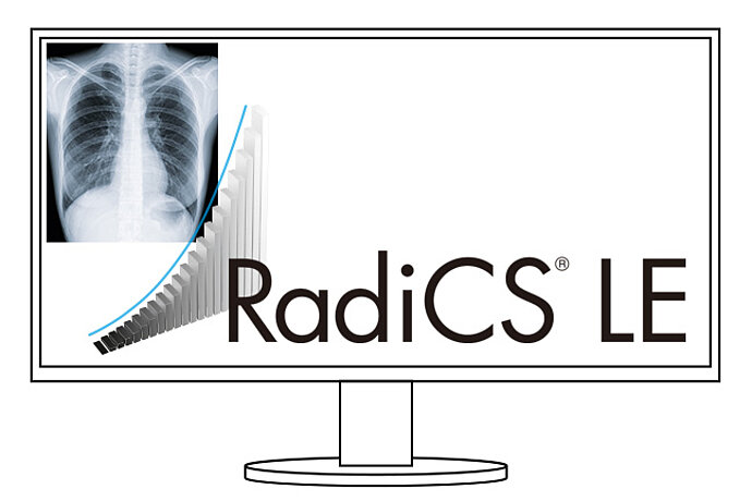
RadiCS LE quality control software calibrates EIZO RadiForce monitors and manages the calibration data.
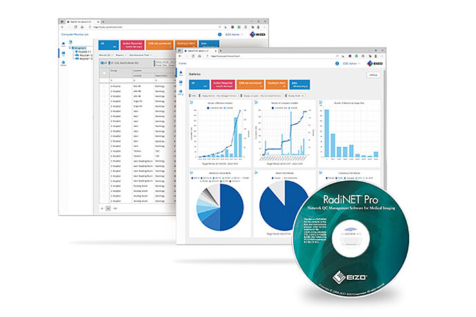
EIZO software for network-based quality management in large facilities – with remote functionality for monitors
ColorNavigator Network enables centralized quality assurance for ColorEdge monitors.
The Screen InStyle Server application allows system administrators to manage and control monitors and PCs connected to the network.
10 of 10 products are shown